We aimed to test the possible differences in gonial angle values between a Medieval sample and a contemporary sample because literature suggests that modern skulls tend to have larger gonial angles. We analyzed the gonial angle values in a Medieval sample (n=69) and a current sample (20th century sample; n=146). We found that current gonial angle values were 3.6º (CI95% 2.2-4.9) larger than the Medieval angle values (p<0.001). No significant differences between the right and left angle values in both the Medieval (p=0.131) and current sample (p=0.120) were observed. The right angle values of the current sample were 3.6º larger (CI95% 1.9-5.3) than the medieval right angle values while the left angle values of the current sample were 3.5º larger (CI95% 1.9-5.2) than the Medieval left angle values. Our research suggests that the present population have larger angle values than the Medieval population.
Gonial angle measures in Medieval and contemporary skeletons
Borja Faus-Valero1, Susanna Llidó-Torrent1, Marcos Miquel-Feutch1, Laura Quiles-Guiñau1, Marcelino Perez-Bermejo1, Shahed Nalla1,2, Juan A. Sanchis-Gimeno1
1 GIAVAL Research Group, Department of Anatomy and Human Embryology, University of Valencia, Faculty of Medicine, Avda. Blasco Ibanez 15, E46010 - Valencia, Spain
2 Department of Human Anatomy and Physiology, Faculty of Health Sciences, University of Johannesburg, Auckland Park, 2006, South Africa
SUMMARY
Sign up or Login
INTRODUCTION
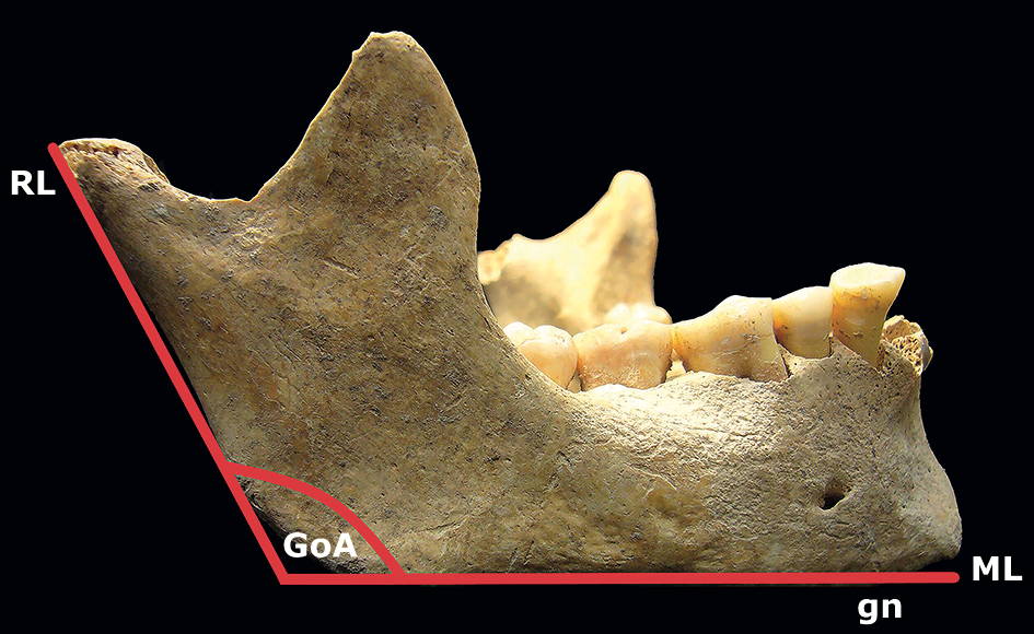
The gonial (mandibular) angle is the angle of the mandible (Fig. 1) formed between the ramus line and the mandibular line as viewed from the lateral aspect of the mandible. The ramus line is the tangent to the posterior border of the mandible, and the mandibular line is the lower border of the mandible through the gnathion (Ohm and Silness, 1999; Dhara et al., 2019).
The gonial angle values have been studied in present day populations (Dutra et al., 2004; Uthman, 2007; Upadhyay et al., 2012; Chole et al., 2013; Bhardwaj et al., 2014; Leversha et al., 2016; Larrazabal-Moron and Sanchis-Gimeno, 2018). Questions about factors that influence the morphology and morphometry of the gonial angle is the focus of research in the scientific community. In this context, different researchers (Varrella, 1990; Luther, 1993; Kaifu, 1997; Gungor et al., 2007; Hayashi et al., 2011; Rando et al., 2014; Toro-Ibacache et al., 2016; Toro-Ibacache et al., 2019) have presented contradictory results after analyzing the jaw size and shape by means of traditional morphometry measurements and geometric morphometrics in order to determine possible morphological changes of the jaw during past centuries, the presence of sexual dimorphism, and the possibility of asymmetry in gonial angle values.
One of the most controversial aspects is the possible larger gonial angle values in contemporary subjects when compared to past subjects. Some authors have suggested that modern skulls tend to have larger gonial angles (Varrella, 1990; Luther, 1993; Rando et al., 2014) due to a modern diet of softer foods (Rando et al., 2014) in comparison to the coarser pre-industrialized diet that required more vigorous masticatory activity (Varrella, 1990). The possible angle changes in line with research that has revealed that mandibular shape varies as a function of the forces applied to it by the temporalis and masseter muscles (Sella-Tunis et al., 2018). However, Gungor et al. (2007) compared the gonial angle values of present subjects with skeletal series from different Anatolian populations to investigate possible changes since the Mesolithic period, and concluded that there is no clear evidence in the change of gonial degree angle in the passage of time.
The study of medieval and contemporary skeletal remains will allow us to answer some of the related research questions mentioned. Following on from this, we aimed to test the possible differences in gonial angle values between medieval and contemporary mandibles in order to test the hypothesis of an increase of the gonial angle values during the past centuries. Secondly, we also aimed to analyze the possible differences in left and right angles and the sexual dimorphism of gonial angle values.
MATERIALS AND METHODS
We carried out an observational transversal study that analyzed the gonial angle values in a medieval sample and a twentieth-century sample. This study was approved by the institutional review board of the University of Valencia (reference number: H1491206464387).
Inclusion criteria of the mandibles in the study are that each specimen of both sample populations required to be of a Caucasian adult, with an age at death ranging from 20 to 50 years old, and have at least one quarter of the dentition presenting teeth numbers 35, 36, 37, 45, 46 and 47. In addition to the above inclusion criteria, the twentieth-century mandibles were obtained from skeletons of subjects who had passed away before 1970 in order to avoid the possible effect of current orthodontic treatments on mandibular morphometric values (Furquim et al., 2018).
The medieval sample was composed of 208 skeletons discovered during the restoration works at a Medieval Muslim burial site of L’Alcudiola (Favara, Valencia, Spain) (Fig. 2). L’Alcudiola Necropolis lies 6 km away from the Mediterranean coast of Spain, around 49 km south of the modern city of Valencia. The human remains found were dated between the eleventh and fourteenth centuries, and after application of the inclusion criteria, 69 of the 208 mandibles (33.2%) found were used in the study.
Due to the lack of availability of old registers on age-at-death of each individual skeleton, the age and sex of said skeletons was determined by evaluation of the metamorphosis at the sternal extremity of the rib, thyroid cartilage ossification, cranial bone synostosis and pubic symphysis morphology (Burns, 2008). The twentieth-century skeletal mandibles (n=146; 100%) with known sex and age at death were obtained from the Raymond A. Dart Collection of Human Skeletons (Dart Collection) housed in the School of Anatomical Sciences, University of the Witwatersrand Medical School, Johannesburg, South Africa. Table 1 presents the demographic characteristics of the samples analyzed.
Measurement of the gonial angles was carried out by means of a conventional mandibulometer. The gonial angle value used was the mean of three consecutive measurements taken on the same day by the same researcher. We also analyzed the sexual dimorphism and the potential differences in the left and the right gonial angle values.
Data were entered and stored in an MS-Excel file, and then transferred to SPSS v.23 software (SPSS Inc., Chicago, IL, USA) for statistical analysis. Continuous variables were presented as mean ± standard deviation. Categorical variables were expressed by count and percentage. Normality of the data distribution was determined by using the Kolmogorov-Smirnov test. Difference between proportions was evaluated utilizing the z-test. Differences between means were analyzed with the paired or unpaired Student t-test as required in the case of two means. Although angles are not meant to be compared using linear statistics, we used a t-test because the differences were within a very small angle range (<10º). Two-sided p<0.05 was considered to be statistically significant. Intraclass correlation coefficient (ICC) estimates and their 95% confident intervals (CI) were calculated based on a 2-way mixed-effects model. ICC values less than 0.5 are considered to be indicative of poor reliability, values between 0.5 and 0.75 indicate moderate reliability, values between 0.75 and 0.9 indicate good reliability, and values greater than 0.90 indicate excellent reliability (Koo and Li, 2016).
RESULTS
In order to evaluate the reliability of our measurements, the gonial angle was measured thrice by the same researcher, and the ICC among the 3 different measurements was calculated. The ICC used to assess measurement accuracy for the gonial angle measurements was 0.995, with a CI95% from 0.991 to 0.997 (F= 193.38, p<0.001), which reflects an excellent reliability. In addition, we also evaluated the results obtained by two different researchers who measured the gonial angles, and we found that the ICC was 0.992 with a CI95% from 0.984 to 0.996 (F= 120.37, p<0.001) which also reflects an excellent reliability.
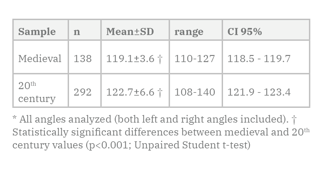
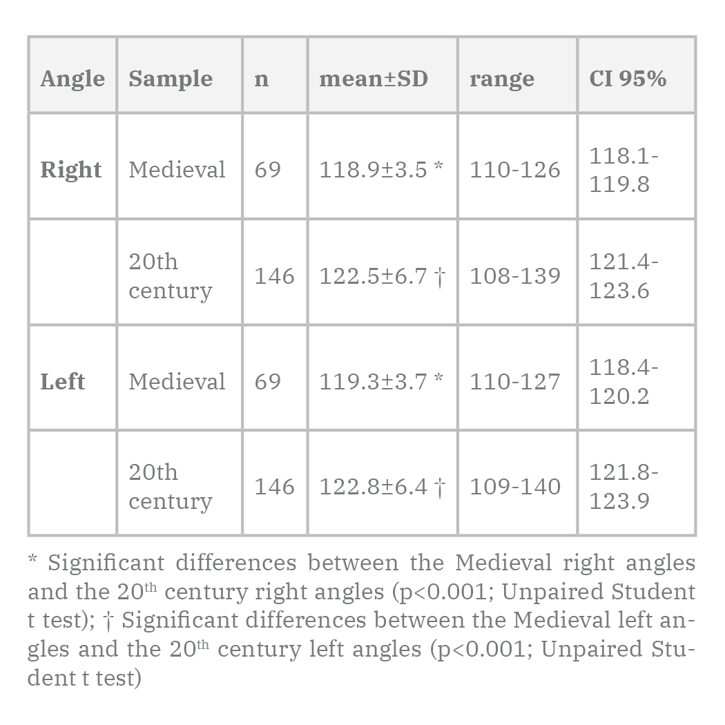
Table 2 presents the gonial angle results obtained in the samples analyzed. We found that the 20th century values were 3.6º (CI95% 2.2-4.9) larger than the Medieval values (p<0.001; Unpaired Student t-test).
Table 3 reveals no significant differences between the right and the left angle values in the medieval (p=0.131; Paired Student t-test) and twentieth-century samples (p=0.120; Paired Student t-test). Nevertheless, we found significant differences when comparing the right angles values of the medieval sample and the right angles values of the twentieth-century sample (p<0.001; Unpaired Student t test). The twentieth-century right angle values were 3.6º larger (CI95% 1.9-5.3) than the medieval right angles values. We found similar results for the left angles (p<0.001; Unpaired Student t-test): the twentieth-century left angle values were 3.5º larger (CI95% 1.9-5.2) than the medieval left angle values.
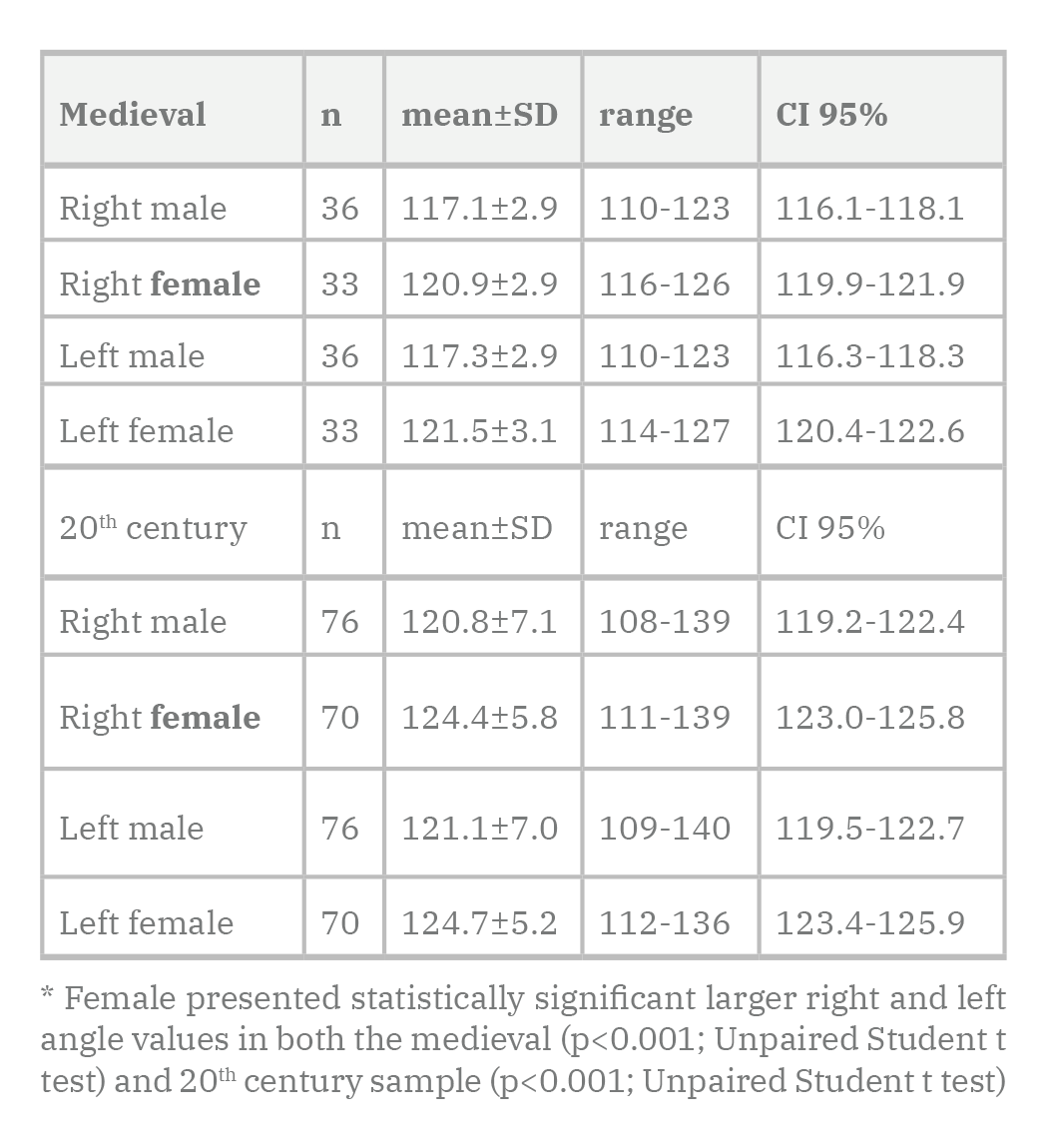
The differences in gonial angle values between females and males are presented in Table 4. Analysis of the medieval sample revealed significant differences between the right values of female and male (p<0.001; Unpaired Student t test): female right angle values were 3.8º larger than male right values (CI95% 2.4-5.2); we also found significant differences between sexes in the left angle values (p<0.001; Unpaired Student t-test), female having left angle values 4.2º larger than male (CI95% 2.8-5.7). The twentieth-century females had right angle values 3.6º larger than that presented by males (p<0.001; Unpaired Student t-test; CI95% 1.4-5.7) while female left values were 3.5º larger than those presented by males (p<0.001; Unpaired Student t-test; CI95% 1.5-5.6).
DISCUSSION
We have carried out a study in two different skeletal samples of subjects aged 20 to 50 years old at death. We measured the gonial angles of adult subjects, and none younger than 20 years old, because the angle values show a continuous decrease until the age of 21 years when it stops changing (Larrazabal-Moron and Sanchis-Gimeno, 2018).
Focusing on the main objective of our research, we observed larger gonial angle values in the twentieth-century sample. In this context, it must be noted that we analyzed skeletons of subjects that passed away before 1970 in order to avoid the effect of current orthodontic treatments in mandibular morphometric values (Furquim et al. 2018), and that the ICC analysis reflected an excellent reliability of the measurements carried out.
Rando et al. (2014) analyzed two skeletal samples from the late medieval period (years 1050-1550) and the post-medieval period (years 1550-1850), and found larger gonial angle values in the post-medieval period sample. Luther (1993) found larger gonial angle values in modern mandibles when compared to medieval mandibles. Our results concur with these authors, as our twentieth-century mandibles present larger gonial angle values than the medieval mandibles.
Similar results were presented by Varrella (1990) as the smaller gonial angles found in fifteenth- and sixteenth-century skulls compared to contemporary; Varrella (1990) suggested that the observed changes in gonial angle values were related to the differences in intensity of chewing between the samples, possibly due to changes in diet (with the present diet being softer), but also concluded that masticatory activity, in addition to breathing, is an important factor in the determination of facial growth direction. Furthermore, Toro-Ibacache et al., (2019) in their study of current and past populations based on the intensity of masticatory loads, found that modern urban subjects with lower intensity of masticatory loads tend to have more gracile features, as well as wider mandibular angles. However, they also indicated that the large variation in the mandible shape of modern subjects may be affected by genetics and nutrition. It must be noted that malnutrition related to social status cannot be factored in a skeletal study undertaken on past skeletal remains like these, as the material properties of food items cannot be measured in extinct populations (Toro-Ibacache et al., 2019), and because malnutrition is a factor that affects the appearance of the jaw (Suazo-Galdames et al., 2008). Thus, Hayashi et al. (2012) recommended identification of the social class of a subject prior to a physical anthropology comparison or examination. The results of their geometric morphometrics analysis revealed a clear difference in craniomandibular shape between the early modern (Edo) Japanese (seventeenth- to nineteenth-century) group, which presented an obtuse gonial angle, and the contemporary Japanese group. Possibly these factors (social and nutrition status), also with the lack of samples from different periods, may be the reason of the non-significant differences between mean gonial angle degrees between late Byzantine, medieval and the contemporary periods observed in the study done in Anatolian populations by Gungor et al., (2007).
Luther (1993) and Rando et al. (2014) also suggested that dietary differences were likely to be major explanatory factors of the differences between medieval and modern mandibles. The daily diet of medieval populations similar to the one studied by us has been analyzed before (Alexander et al., 2015; Guede et al., 2017). These studies have revealed that the diet was based on cereals, pulses, lamb, poultry, fish, fruit, honey, eggs, milk and cheese, which was similar to twentieth- and twenty-first-century daily diet comprising plant foods, olive oil, fish, poultry, red meat, fresh fruit, refined white maize meal, bran, sugar, and vegetables (Abramson et al., 1960; Widmer et al., 2015). Nonetheless, the current human diet is softer than pre-industrialized diet because of food processing technologies (Rando et al., 2014) may imply that less force is needed to be applied by the temporalis and masseter muscles during biting, being a possible explanation for the current larger gonial angle values, because of the relationship between the force input and the cranial skeletal deformation during biting (Toro-Ibacache et al., 2016). This may be one possible cause of the larger gonial angle values observed in the twentieth-century sample. Moreover, it is known that the mandible is considered to be a bone whose morphology is adaptable (Nicholson and Harvati, 2006; Smith, 2009). Kaifu (1997) found a human mandibular reduction in the Japanese remains between the Kofun and the Edo periods that may be related to a reduction of chewing stress. Similarly, May et al. (2018) and Pokhojaev et al. (2019) suggested that changes in mandibular size and orientation during the terminal Pleistocene to Holocene can be explained by a reduction in the biomechanical demands of the masticatory system.
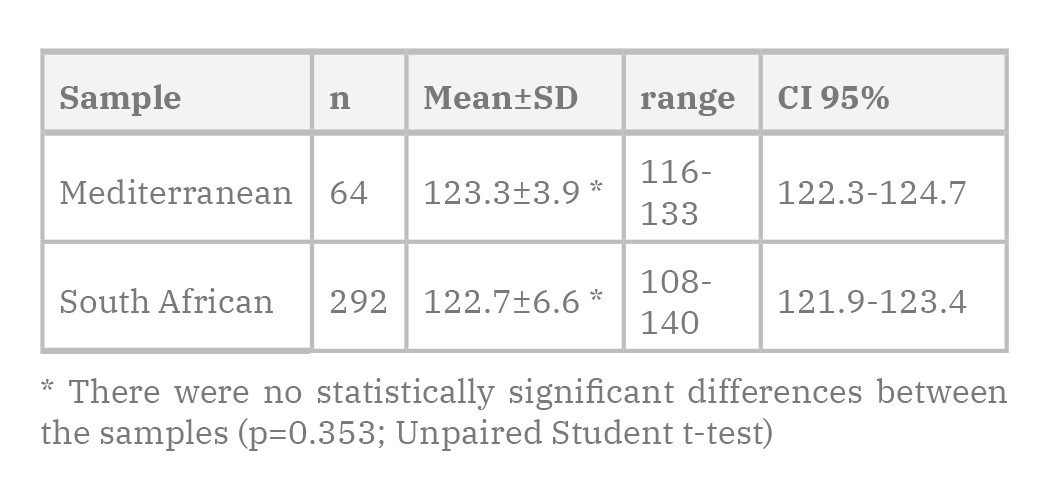
All this raises a question about the comparability of the samples we studied, as the modern sample was obtained from a South African skeletal collection housed in a modern urban city, whereas the Medieval sample was composed of Mediterranean skeletons from a small rural location. It could be argued that both samples were not comparable due to a number of factors, for example, possible different social status, sample composition, geographical differences as well as ancestry and genetic differences. Therefore, we have compared the twentieth-century gonial angles from the Dart collection we used with the gonial angles of a twentieth-century Mediterranean skeletal sample from our lab, and we found no significant differences between both the twentieth-century Mediterranean and South African samples (Table 5). The gonial angles were also significantly larger in the contemporary Mediterranean sample than in the medieval Mediterranean sample (p<0.001; Unpaired Student t-test).
Regarding asymmetry and sexual dimorphism, we found no significant differences between the right and the left angle values in the medieval and twentieth-century samples, but females presented statistically significant larger angle values in both the medieval and twentieth-century samples, our samples being well balanced between sexes. Gungor et al. (2007) found no asymmetrical difference between the right and left gonial angle degrees of individuals belonging to the same sex, but females have larger gonial angles, which leads to concluding that mandibular gonial angle showed sexual dimorphism with female having higher values starting from the early settlement times in Anatolia through time. In addition, Hayashi et al. (2012) found morphological differences between early modern (Edo) Japanese women and contemporary Japanese women using human female remains, and although they noted that the reason of their results remained unclear, they related the results possibly to both environmental changes and genetic variation over the past 100 years. Similarly, Katz et al. (2017) also proposed that genetic mechanisms are involved in skull shape. In this context, Coquerelle et al. (2011) highlight that sexual dimorphism in the mandible is closely related to the soft tissues of the oral cavity, because these are a major component of facial growth.
Anatomical analysis of ancient and present skeletons allows for reverse translational research to be undertaken. For example, the detected increased gonial angle values in present subjects, when compared to skeletons of populations who lived several centuries ago, may provide information about possible clinical symptoms and/or pathologies that current people may indicate. In this context, our results may have future research implications, as it appears that there is a positive correlation between high gonial angle values and the presence of an angle fractures (Panneerselvam et al., 2017; Bereznyak-Elias et al., 2018; Dhara et al., 2019), which are the most common mandibular fractures (Aleysson et al., 2008). Subjects with smaller gonial angles (as observed in our medieval sample) are assumed to have greater muscle activity and thus greater cortical bone thickness (Jonasson and Kiliaridis, 2004; Osato et al., 2012), while subjects with larger gonial angle values (as observed in our twentieth-century sample) that generate relatively decreased bite forces or masticatory loads will present decreased cortical bone thickness (Panneerselvam et al., 2017). Thus, results obtained in this study may be complemented with further skeletal research in order to determine whether modern skeletons present a high incidence of angle fractures than historical skeletons.
In summary, our study suggests that contemporary skeletons present larger gonial angle values than Medieval skeletons maybe due to a current softer diet. Further research is required in order to determine the possible effect on mandibular shape and gonial angle values of factors such as social status, nutrition, geographical differences, ancestry, and genetic differences.
ACKNOWLEDGEMENTS
The authors wish to thank Miss Manuela Raga y Rubio, Head of Archeological Intervention in L’Alcudiola, for her assistance in different phases of this research work.
Related articles
ABRAMSON JH, SLOME C, WARD NT (1960) Diet and health of a group of African agricultural workers in South Africa. Am J Clin Nutr, 8: 875-884.
ALEXANDER MM, GERRARD CM, GUTIÉRREZ A, MILLARD AR (2015) Diet, society, and economy in late medieval Spain: stable isotope evidence from Muslims and Christians from Gandía, Valencia. Am J Phys Anthropol, 156: 263-273.
ALEYSSON PO, ABUABARA A, PASSERI LA (2008) Analysis of 115 mandibular angle fractures. J Oral Maxillofac Surg, 66: 73-76.
BEREZNYAK ELIAS Y, SHILO D, EMODI O, NOY D, RACHMIEL A (2018) The Relation Between Morphometric Features and Susceptibility to Mandibular Angle Fractures. J Craniofac Surg, 29: e663-e665.
BHARDWAJ D, KUMAR JS, MOHAN V (2014) Radiographic evaluation of mandible to predict the gender and age. J Clin Diagn Res, 8: 66-69.
BURNS KR (2008) Forensic Anthropology Manual. Edicions Bellaterra SL, Barcelona.
CHOLE RH, PATIL RN, BALSARAF CHOLE S, GONDIVKAR S, GADBAIL AR, YUWANATI MB (2013) Association of mandible anatomy with age, gender, and dental status: a radiographic study. ISRN Radiol, 2013: 453763.
COQUERELLE M, BOOKSTEIN FL, BRAGA J, HALAZONETIS DJ, WEBER GW, MITTEROECKER P (2011) Sexual dimorphism of the human mandible and its association with dental development. Am J Phys Anthropol, 145: 192-202.
DHARA V, KAMATH AT, VINEETHA R (2019) The influence of the mandibular gonial angle on the occurrence of mandibular angle fracture. Dent Traumatol, 35: 188-193.
DUTRA V, YANG J, DEVLIN H, SUSIN C (2004) Mandibular bone remodelling in adults: evaluation of panoramic radiographs. Dentomaxillofac Radiol, 33: 323-328.
FURQUIM BD, JANSON G, COPE LCC, FREITAS KMS, HENRIQUES JFC (2018) Comparative effects of the mandibular protraction appliance in adolescents and adults. Dental Press J Orthod, 23: 63-72.
GUEDE I, ORTEGA LA, ZULUAGA MC, ALONSO-OLAZABAL A, MURELAGA X, PINA M, GUTIERREZ FJ, IACUMIN P (2017) Isotope analyses to explore diet and mobility in a medieval Muslim population at Tauste (NE Spain). PLoS One, 12: e0176572.
GUNGOR K, SAGIR M, OZER I (2007) Evaluation of the gonial angle in the Anatolian populations: from past to present. Coll Antropol, 31: 375-378.
HAYASHI K, SAITOH S, MIZOGUCHI I (2011) Morphological analysis of the skeletal remains of Japanese females from the Ikenohata-Shichikencho site. Eur J Orthod, 34: 575-581.
JONASSON G, KILIARIDIS S (2004) The association between the masseter muscle, the mandibular alveolar bone mass and thickness in dentate women. Arch Oral Biol, 49: 1001-1006.
KAIFU Y (1997) Changes in mandibular morphology from the Jomon to modern periods in eastern Japan. Am J Phys Anthropol, 104: 227-243.
KATZ DC, GROTE MN, WEAVER TD (2017) Changes in human skull morphology across the agricultural transition are consistent with softer diets in preindustrial farming groups. PNAS, 114: 9050-9055.
KOO TK, LI MY (2016) A guideline of selecting and reporting intraclass correlation coefficients for reliability research. J Chiropr Med, 15: 155-163.
LARRAZABAL-MORON C, SANCHIS-GIMENO JA (2018) Gonial angle growth patterns according to age and gender. Ann Anat, 215: 93-96.
LEVERSHA J, MCKEOUGH G, MYRTEZA A, SKJELLRUP-WAKEFILED H, WELSH J, SHOLAPURKAR A (2016) Age and gender correlation of gonial angle, ramus height and bigonial width in dentate subjects in a dental school in Far North Queensland. J Clin Exp Dent, 8: e49-54.
LUTHER F (1993) A cephalometric comparison of medieval skulls with a modern population. Eur J Orthod, 15: 315-325.
MAY H, SELLA-TUNIS T, POKHOJAEVA A, PELED D, SARIG R (2018) Changes in mandible characteristics during the terminal Pleistocene to Holocene Levant and their association with dietary habits. J Archaeol Sci Rep, 22: 413-419.
NICHOLSON E, HARVATI K (2016) Quantitative analysis of human mandibular shape using three-dimensional geometric morphometrics. Am J Phys Anthropol, 131: 368-383.
OHM E, SILNESS J (1999) Size of the mandibular jaw angle related to age, tooth retention and gender. J Oral Rehabil, 26: 883-891.
OSATO S, KUROYAMA I, NAKAJIMA S, OGAWA T, MISAKI K (2012) Differences in 5 anatomic parameters of mandibular body morphology by gonial angle size in dentulous Japanese subjects. Ann Anat, 194: 446-451.
PANNEERSELVAM E, PRASAD PJ, BALASUBRAMANIAM S, SOMASUNDARAM S, RAJA KV, SRINIVASAN D (2017) The influence of the mandibular gonial angle on the incidence of mandibular angle fracture-a radiomorphometric study. J Oral Maxillofac Surg, 75: 153-159.
POKHOJAEV A, AVNI H, SELLA-TUNIS T, SARIG R, MAY H (2019) Changes in human mandibular shape during the Terminal Pleistocene-Holocene Levant. Sci Rep, 9: 8799.
RANDO C, HILLSON S, ANTOINE D (2014) Changes in mandibular dimensions during the medieval to post-medieval transition in London: a possible response to decreased masticatory load. Arch Oral Biol, 59: 73-81.
SELLA-TUNIS T, POKHOJAEV A, SARIG R, O'HIGGINS P, MAY H (2018) Human mandibular shape is associated with masticatory muscle force. Sci Rep, 8: 6042.
SMITH HF (2009) Which cranial regions reflect molecular distances reliably in humans? Evidence from three-dimensional morphology. Am J Hum Biol, 21: 36-47.
SUAZO-GALDAMES IC, ZAVANDO-MATAMALA DA, LUIZ-SMITH R (2008) Evaluating accuracy and precision in morphologic traits for sexual dimorphism in malnutrition human skull: a comparative study. Int J Morphol, 26: 877-881.
TORO-IBACACHE V, UGARTE F, MORALES C, EYQUEM A, AGUILERA J, ASTUDILLO W (2019) Dental malocclusions are not just about small and weak bones: assessing the morphology of the mandible with cross-section analysis and geometric morphometrics. Clin Oral Investig, 23: 3479-3490.
TORO-IBACACHE V, ZAPATA MUÑOZ V, O'HIGGINS P (2016) The relationship between skull morphology, masticatory muscle force and cranial skeletal deformation during biting. Ann Anat, 203: 59-68.
UPADHYAY RB, UPADHYAY J, AGRAWAL P, RAO NN (2012) Analysis of gonial angle in relation to age, gender, and dentition status by radiological and anthropometric methods. J Forensic Dent Sci, 4: 29-33.
UTHMAN AT (2007) Retromolar space analysis in relation to selected linear and angular measurements for an Iraqi sample. Oral Surg Oral Med Oral Pathol Oral Radiol Endod, 104: e76-82.
VARRELA J (1990) Effects of attritive diet on craniofacial morphology: a cephalometric analysis of a Finnish skull sample. Eur J Orthod, 12: 219-223.
WIDMER RJ, FLAMMER AJ, LERMAN LO, LERMAN A (2015) The Mediterranean diet, its components, and cardiovascular disease. Am J Med, 128: 229-238.

